A7 · Clinical conditions
Bone augmentation before a dental implant: when it's needed
Bone augmentation rebuilds ridge volume when there is not enough structure to seat an implant safely. This article explains when grafting is necessary, compares the four material families — autograft, allograft, xenograft, alloplast — across healing time and indication, walks through block, particulate, and guided bone regeneration formats, and shows why alveolar ridge preservation at extraction cuts later resorption by roughly half.
- Published
- 2026-05-20
- Reading time
- 9 minutes
- Author
- Prof. Robert Ćelić
- Section
- Clinical conditions
At our specialist clinic for dental implants in Zagreb, the third most common preparatory step we discuss with Swedish patients is bone augmentation — what most patients have heard about as "bone graft" and what most are quietly worried about because it sounds invasive. The honest version: there are four graft material families, the differences between them matter, and modern bone augmentation is a smaller deal than the word "graft" implies.
What is bone augmentation, and when is it necessary?
Bone augmentation — benuppbyggnad in Swedish — is the surgical addition of bone or bone-substitute material to a deficient alveolar ridge. The goal is to give an implant enough bone volume to anchor in.
A quick note on terminology. Swedish dental practice uses benuppbyggnad for any graft type. Bentransplantation is technically a subset: it specifically means transplanting bone from another site (autologous bone). When the graft is allograft, xenograft, or synthetic, "transplantation" is the wrong word. We use benuppbyggnad consistently because it covers all four material families correctly.
When is it actually needed? The honest answer is: in roughly a quarter of implant cases, depending on what happened after the original tooth was lost. The main triggers are:
- · Tooth loss without immediate post-extraction socket preservation — ridge width typically loses about 30–50% in the first 12 months, with most of the loss in months three to six. Cumulative loss of 40–60% is more typical over the first 1–3 years (Schropp 2003; Tan et al. systematic review).
- · Long-term edentulism — years without teeth in a region accelerates the loss.
- · Trauma or congenital absence.
- · A failed previous implant requiring re-grafting.
- · Sinus floor proximity in the upper posterior maxilla — covered separately in our sinus lift before a dental implant article.
A prevention angle worth knowing. If the tooth has not yet been extracted but is condemned, an immediate post-extraction graft can be placed in the socket. This is called alveolar ridge preservation, or ARP. It reduces ridge dimensional loss in the first 3–6 months by roughly half (Jung 2013; Avila-Ortiz 2014). ARP does not fully prevent resorption; it mitigates it. For patients still considering extraction timing, raising the option early can avoid a larger augmentation later.
The clinical reality is that bone augmentation has become routine over the last twenty years. The Aghaloo & Moy 2007 systematic review established it as predictable across techniques. The question is no longer whether augmentation works. It is which technique and material are right for your specific defect.
The four graft material families — and what each is good for
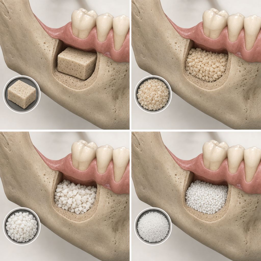
1. Autologous bone — your own bone, harvested from another site.
- · Source: usually the mandibular ramus or chin (intraoral); rarely the iliac crest for very large reconstructions
- · Pros: gold standard for bone biology; contains living osteogenic cells; fastest integration (3–4 months for small-to-moderate intraoral grafts; 4–6 months for larger reconstructions)
- · Cons: a second surgical site means donor-site morbidity; limited volume available; longer operative time
- · Best for: small to moderate vertical augmentations with healthy donor sites elsewhere
2. Allograft — processed human donor bone.
- · Source: tissue banks under FDA / EU regulation
- · Pros: no donor-site surgery; unlimited supply; well-characterised integration
- · Cons: slower integration (4–6 months); a theoretical (but extremely low) disease-transmission risk; some patients prefer not to use human donor tissue for personal reasons
- · Best for: moderate-to-large defects where avoiding a second surgical site matters
3. Xenograft — animal-derived bone, almost always bovine. Bio-Oss is the canonical example.
- · Source: bovine bone, fully de-proteinized and sterilized
- · Pros: unlimited supply; excellent long-term volume stability (the material resorbs very slowly, acting as a long-term scaffold); over 30 years of clinical data
- · Cons: slower biological integration (6–9 months); some patients prefer not to use animal-derived material for dietary or religious reasons
- · Best for: sinus lifts, large augmentations where long-term volume preservation matters most
4. Synthetic — lab-manufactured (β-TCP, hydroxyapatite, biphasic calcium phosphate, bioactive glass).
- · Source: biocompatible ceramic and glass materials manufactured to defined specifications
- · Pros: no biological-source concerns; predictable behaviour; the lowest cost of the four
- · Cons: osteoconductive only (a passive scaffold), not osteoinductive; less long-term clinical data than xenograft
- · Integration nuance: β-TCP-based materials typically ready at 3–6 months; HA-rich or dense ceramic synthetics often require 6–9 months and leave long-term remnants
- · Best for: smaller defects, or patients who prefer not to use human or animal grafts
An emerging fifth option worth mentioning. Tooth-derived autograft uses the patient's own extracted teeth — cleaned, demineralized, and chairside-processed — as graft material. Limited but encouraging clinical evidence (Kim 2017 and follow-ups) suggests new bone formation comparable to xenograft and allograft at 4–6 months, with no donor-site morbidity. Not yet standard of care, but a real option in selected centres for patients who happen to be extracting a tooth at the same visit.
The right choice for your specific case depends on the defect geometry, the volume needed, your overall health, and your personal preferences about graft source. We make that decision at consultation based on CBCT imaging and clinical exam — not from a brochure. For broader context on the implant pathway, see our dental implants in Croatia overview. Technique selection across material types is reviewed in the Esposito et al. 2009 Cochrane-method review.
How the procedure works — block grafts, particulate, and GBR
There are three real surgical formats. Each fits a different defect type.
Block graft.
- · A solid piece of bone (usually autologous, harvested from the ramus or chin) is fixed to the host site with mini-screws
- · Indicated for large three-dimensional defects where particulate would not hold shape
- · 4–6 months of integration before implant placement
- · Higher technique demand, longer recovery
- · Implant survival in block-augmented sites: 93–100% at 1–5 years (Starch-Jensen 2016 systematic review)
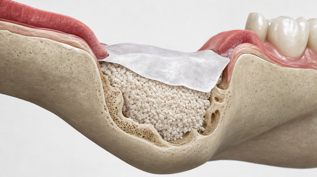
Particulate graft.
- · Granular material (allograft, xenograft, synthetic, or a mix with autologous chips) is packed into the defect
- · Almost always combined with a barrier membrane — see GBR below
- · Indicated for moderate defects, sinus lifts, and socket preservation
- · 4–9 months of integration depending on material
Guided bone regeneration (GBR) — the modern standard for most augmentations.
GBR adds a barrier membrane over the particulate graft. The membrane keeps soft tissue out of the graft space, so only bone-forming cells reach the defect. The contemporary consensus is:
- · For horizontal defects — a resorbable collagen membrane with particulate graft is the standard. Fewer complications than non-resorbable options. Implant survival in horizontally augmented ridges is 95–100% at 1–5 years (Urban 2018 systematic review).
- · For vertical or complex defects — non-resorbable titanium-reinforced PTFE or, increasingly, a CAD/CAM custom titanium mesh holds space for larger vertical gains (typically 4–7 mm). Implant survival is 92–98%; the trade-off is a higher mesh-exposure rate of 20–30%, usually manageable without losing the entire graft.
- · For socket preservation specifically — small-pore-size dPTFE is favoured in some protocols because it resists bacterial penetration and can be left partially exposed.
A real modern trend worth knowing. The contemporary direction is to prefer horizontal GBR and ARP over large vertical onlay grafts whenever the prosthetic plan permits. Narrow-diameter implants in selected cases of ridges as narrow as ~4.5 mm can avoid extensive horizontal augmentation entirely (Donos et al. 2023 — 10-year outcomes published). Less invasive, faster recovery, fewer complications. Not always possible, but always worth checking on the CBCT before committing to a major graft.
Honest framing per voice guide §5:
There is no universal best technique. The right approach depends on defect size, defect geometry, your overall health, and the implant timeline. A surgeon who recommends the same approach for every patient should be a warning sign.
Recovery, integration time, and what affects success
Recovery from a well-performed augmentation is similar to recovery from a wisdom tooth extraction. Most Swedish patients we treat describe days three to four as the worst, with significant improvement by day seven.
Typical timeline:
- · Day 0: procedure under local anaesthesia, with optional sedation. Patient returns to hotel or home the same day.
- · Days 1–7: mild-to-moderate swelling, soft diet, antibiotic and anti-inflammatory regimen. Avoid pressure on the surgical site.
- · Weeks 2–4: sutures dissolve or are removed at the week 1–2 visit. Most discomfort resolves.
- · Months 3–9: the graft integrates. A CBCT at 4–9 months (timing depends on material and volume) confirms readiness for implant placement.
Patient-side factors that affect success. Smoking is the single biggest modifiable risk. The Chrcanovic 2016 meta-analysis gives an odds ratio of 2.17 — smokers face roughly double the implant failure risk of non-smokers. For grafts specifically, smoking elevates complication rates 2–3× (membrane exposure, infection, partial graft loss — Kim 2019). The cessation window we recommend is 4–8 weeks pre-op and continued abstinence post-op. This is expert consensus extrapolated from periodontal and general surgical literature rather than a hard RCT-locked number, but the direction of the evidence is unambiguous.
Diabetes control matters too. Well-controlled diabetes (HbA1c below 7.5% — the modern threshold, covered in our healing process explainer — is not an exclusion. Poorly controlled diabetes meaningfully impairs graft integration. Oral hygiene at the surgical site and post-op medication compliance round out the patient-side picture.
Clinician-side factors. Atraumatic flap design and tension-free primary closure are the technique fundamentals. Graft material must match the defect. Membrane perforation during placement leads to graft exposure and resorption.
Material-side factors. Integration speed varies by material as covered above. Long-term volume stability is best with xenograft, moderate with synthetic and allograft, and excellent (but resorption-prone) with autograft.
Complication-profile transparency. This matters for informed consent. Horizontal GBR and ARP carry complication rates under 10–15%, mostly minor (temporary swelling, slight wound dehiscence). Vertical GBR and block grafts carry 20–30% early complication rates — typically membrane exposure or partial graft loss. The complications are usually manageable without losing the augmentation, but you should know the rates before signing consent.
A patient-reassurance point worth stating directly. Implants placed in properly augmented bone show similar long-term marginal bone loss to implants in native bone — about 0.5–1.0 mm in the first year and 0.1 mm per year thereafter (Urban 2018). Grafted bone is not inherently inferior. It is bone that needed a head start.
Honest framing per voice guide §5:
Bone augmentation has become routine in modern implant dentistry — but only because the technique, material, and patient selection are correct. A "we'll figure it out when you get here" approach is a warning sign, not a flexible plan.
Bone augmentation is one of those procedures where surgeon experience, technique selection, and material choice compound visibly in long-term outcomes. The peer-reviewed difference between a 90% success rate and a 97% success rate is real, and it is the difference between routine and exceptional clinical practice. If you have been told you need a graft and want a CBCT-based second opinion from a named specialist with a verifiable EDA-Expert credential, Prof. Robert Ćelić, European Expert in Implantology reviews cases personally in a free 30-minute consultation. No deposit, no commitment.
References
Sources referenced.
- Aghaloo TL, Moy PK (2007) — Which hard tissue augmentation techniques are the most successful in furnishing bony support for implant placement? Int J Oral Maxillofac Implants 2007;22(Suppl):49–70. PMID 17542462. External link in H2.1.
- Esposito M, Grusovin MG, Felice P, Karatzopoulos G, Worthington HV, Coulthard P (2009) — The efficacy of horizontal and vertical bone augmentation procedures for dental implants — a Cochrane systematic review. Eur J Oral Implantol 2009;2(3):167–184. PMID 20467591. External link in H2.2.
- Schropp L et al. (2003) — post-extraction ridge dimensional changes (~50% width reduction at 12 months). PMID 14761119. In-text.
- Tan WL et al. — systematic review of post-extraction ridge dimensional changes (up to 3.8 mm horizontal / 1.24 mm vertical loss within 6 months). PMID 17949342. In-text.
- Urban IA et al. (2018) — horizontal GBR systematic review; implant survival 95–100%. PMID 29503825. In-text.
- Starch-Jensen T et al. (2016) — autogenous block grafts systematic review; implant survival 93–100%. PMID 27045421. In-text.
- Jung RE et al. (2013) — alveolar ridge preservation systematic review. PMID 24164305. In-text.
- Avila-Ortiz G et al. (2014) — ARP meta-analysis. PMID 24482353. In-text.
- Chrcanovic BR et al. (2016) — smoking and implant survival meta-analysis (OR 2.17). PMID 27363721. In-text.
- Kim Y et al. (2019) — smoking and bone augmentation complications. PMID 30426625. In-text.
- Donos N et al. (2023) — 10-year outcomes of implants in narrow alveolar ridges. PMID 36930063. In-text.
- Tooth-derived autograft clinical evidence — Kim 2017 series and follow-ups. PMIDs 28960235, 28200368. In-text.
- CAD/CAM custom titanium mesh outcomes — PMIDs 34462223, 33340839. In-text.
Read next
Three pages worth your time.
The Pezo–Ćelić Protocol
Dental implants in Croatia — 20-year written guarantee
The full offer stack, dual guarantees, and surgeon-team accountability detailed.
Named credentials
Prof. Ćelić, European Expert in Implantology
Full Professor at University of Zagreb. BDIZ EDI certified. 916+ citations.
Related article
Osseointegration — how an implant fuses with the jawbone
What grafted bone has to achieve next: locking onto the implant surface through the three phases of osseointegration.
Ready when you are
Want a CBCT-based assessment from a named specialist?
A free 30-minute consultation. Send us your X-rays or current treatment plan. We will review it, give you our honest assessment, and confirm whether you are a candidate. If we do not think we are the right fit for your case, we will say so.
No deposit. No commitment.